
- → Thoughts move at 270 mph, not the speed of light: Nerve signals do not rely on free electrons in metal, but on the physical movement of ions across cellular membranes (Rushton, 1951).
- → The myelin sheath is a biological fiber optic jacket: Composed of lipids and proteins, this specialized insulation prevents charge leakage and accelerates conduction velocity by up to 100-fold (Ford et al., 2015).
- → Nodes of Ranvier act as signal repeaters: Rather than continuous flow, the action potential jumps between nodes, saving thousands of times more metabolic cellular energy (Sengupta et al., 2010).
- → Myelin degradation causes cognitive lag: Inadequate B12, chronic sleep deprivation, and sustained neuroinflammation wear down myelin, prolonging reaction times (Harris & Attwell, 2012).
⚠️ Informational Notice: This article provides general educational information based on peer-reviewed biophysical and neurological literature and does not substitute for clinical medical advice. If you experience persistent numbness, tingling, or neurological symptoms, consult a qualified physician.
In Brief
- Thoughts do not travel at light speed; in our fastest motor fibers, nerve impulses peak at roughly 270 miles per hour (120 meters per second) [1].
- This impressive speed is made possible by the myelin sheath, a multi-layered lipid insulation surrounding nerve fibers (axons) [1, 2].
- Rather than traveling continuously down the wire, impulses jump between Nodes of Ranvier, reducing cellular energy consumption by up to 5,000-fold [3, 4].
- The good news: Supporting and protecting your myelin sheath is directly achievable through targeted nutrition, physical activity, and deep restorative sleep.
Sounds Familiar?
You are driving on a highway at 70 miles per hour in flowing traffic. Suddenly, the brake lights of the car ahead flash bright red.
Your foot lifts off the accelerator and depresses the brake pedal in a split second—around 150 milliseconds. In that intense instant, everything feels instantaneous; your mind whispers, “I reacted at the speed of light.”
Yet in physical reality, none of our conscious thoughts or motor reflexes move anywhere near the speed of light. In the brief moment between your eyes registering photons and your calf muscles engaging, biological fiber optic conduits deep in your nervous system race against time.

1. Why Is Thought Slower Than Light?
In a vacuum, light races at roughly 186,000 miles per second (300,000 km/s). Inside submarine glass fiber cables, laser pulses travel at about two-thirds of that cosmic limit.
In stark contrast, electrical impulses in our most specialized nerve fibers top out at roughly 120 meters per second (about 270 mph / 430 km/h) [1]. In unmyelinated fibers carrying dull pain or temperature, the signal crawls along at walking speed—barely 4 miles per hour [1].
The fundamental reason is physical: our nervous system does not channel free-flowing electrons through solid copper. Nerve signaling is an electro-chemical wave driven by the physical opening and closing of microscopic ion channels, allowing sodium and potassium ions to cross cellular membranes [1, 3].
2. Axons and Myelin: Biological Fiber Optics
Consider the architecture of modern subsea fiber optic cables. At the center lies a hair-thin glass core transmitting photons, surrounded by protective optical cladding and water-resistant protective jackets that prevent light leakage.
The mammalian brain operates on an identical engineering principle. The long signaling projections of our neurons are called axons (nerve transmission cables). Surrounding these biological wires is the myelin sheath (biological insulation jacket), formed by concentric layers of specialized lipids and structural proteins [2].
Without this insulation, electrical charge would bleed across the leaky axon membrane, dissipating the signal long before it could reach target muscles [2]. Myelin minimizes membrane capacitance and boosts electrical resistance, ensuring information reaches its destination without loss [1, 2].

3. Saltatory Conduction: Microscopic Signal Repeaters
In transcontinental optical networks, light signals degrade over distance without periodic inline optical amplifiers (repeaters) to regenerate the wave.
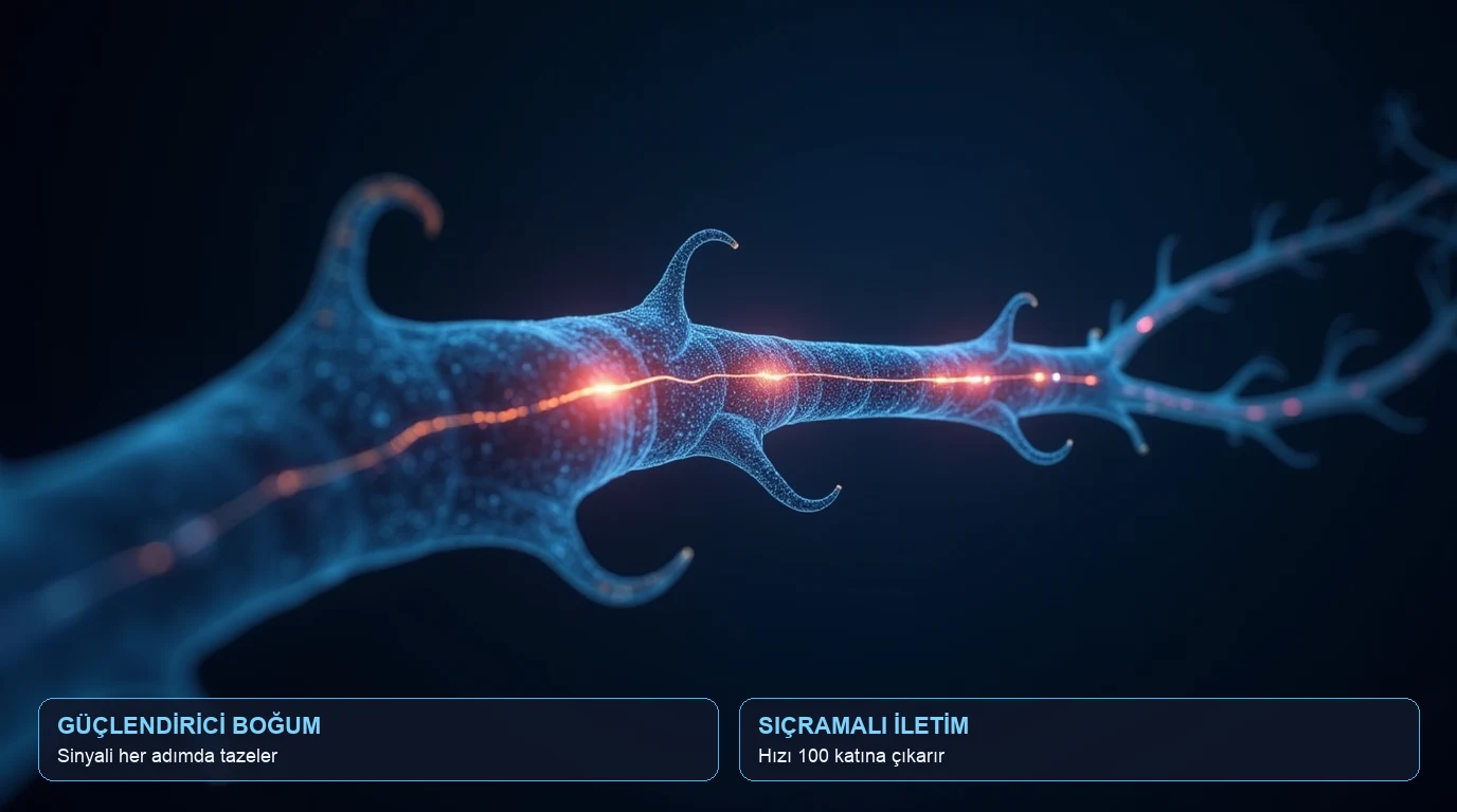
Our biological cables feature identical booster stations placed every millimeter along the axon: the Nodes of Ranvier (signal-regenerating booster junctions) [2].
Myelin does not coat the axon as a seamless sleeve; it leaves tiny exposed gaps. Electrical currents flow almost instantaneously through the insulated segments, and upon hitting each node, clusters of voltage-gated sodium channels open to recharge the action potential to full amplitude [2, 3].
The impulse literally jumps from node to node like a kangaroo. Neurophysiologists term this saltatory conduction (from Latin saltare, to leap) [1, 3]. This jumping mechanism accelerates conduction velocity by up to 100 times compared to unmyelinated fibers of equivalent diameter [2].
4. Massive Metabolic Savings and Skull Volume
What if biology had never invented myelination? In the evolutionary arms race, giant squids achieved rapid jet-propulsion reflexes by evolving unmyelinated axons up to 1 millimeter in diameter [3].
If the human brain relied on unmyelinated cables to match our current reflex and cognitive processing speeds, our nerve fibers would need to be as thick as pencils [3]. In that scenario, our skull would have to be as large as a living room, rendering human bipedal posture impossible [1, 3].
Furthermore, saltatory conduction dramatically slashes the brain’s metabolic budget. Because ATP-consuming sodium-potassium pumps are only active at the tiny nodal gaps rather than across the entire axon surface, cellular energy expenditure during signaling is reduced by up to 5,000-fold [3, 4].

5. Nutrition for Resilient Neural Cables
Between 70% and 80% of dry myelin sheath weight consists of specialized lipids (sphingolipids, cholesterol, galactolipids), with the remainder made of structural scaffolding proteins [4]. The integrity of this insulation depends heavily on nutritional substrates.
- Vitamin B12 and Folate: Indispensable cofactors in the methylation cycles required for myelin synthesis. Deficiency causes subacute combined degeneration, presenting as peripheral tingling, cognitive slowing, and diminished reflexes [4].
- Choline (Egg Yolks): Essential precursor for sphingomyelin and phosphatidylcholine, the foundational lipid bilayers of nerve insulation.
- Omega-3 Fatty Acids (DHA): Maintains structural membrane fluidity, ensuring rapid ion channel gating and resilience against oxidative stress [4].
- Iron: A critical cofactor for oligodendrocyte progenitor cell differentiation and myelinogenesis.

6. How Deep Sleep and Movement Rebuild Myelin
During sustained daily cognitive engagement and chronic stress, neural insulation incurs micro-wear. Fortunately, our physiology features a dedicated nocturnal repair protocol.
- Nocturnal Maintenance (Deep Slow-Wave Sleep): During non-REM deep sleep, the brain’s glymphatic clearance system flushes metabolic debris while genes involved in phospholipid synthesis and remyelination double their transcription rates [4].
- Physical Aerobic Activity: Brisk walking and zone-2 cardiovascular training stimulate brain-derived neurotrophic factor (BDNF), activating oligodendrocyte maturation and the reinforcement of active neural pathways.

What You Can Do: 4 Actionable Steps
- Eat 1 to 2 pasture-raised boiled eggs daily: Egg yolks are nature’s richest source of bioavailable choline and B12 for myelin lipid turnover.
- Incorporate oily fish or walnuts twice weekly: Marine DHA and plant-based ALA support lipid membrane flexibility and prevent conduction attenuation.
- Guard a minimum of 7 hours of nocturnal sleep: Structural myelin remodeling and glymphatic detoxification require unbroken deep sleep cycles.
- Engage in 25–30 minutes of daily aerobic movement: Aerobic conditioning upregulates neurotrophic factors that trigger remyelination. (The simplest, most impactful habit.)
Evidence Quality Appraisal
| Finding | Evidence Level |
|---|---|
| Myelin insulation accelerates conduction velocity up to 100-fold | 🟢 Robust |
| Saltatory conduction reduces action potential ATP consumption by thousands of times | 🟢 Robust |
| B12 and choline deficiency precipitates demyelination and neurological deficits | 🟢 Robust |
| Deep sleep doubles transcription of remyelination genes | 🟡 Emerging |
💡 What Do These Confidence Levels Mean?
- 🟢 Robust: Confirmed across multiple independent human clinical trials, robust electrophysiology, and broad scientific consensus.
- 🟡 Emerging: Promising preliminary human trials, mechanistic animal studies, or specialized pilot cohorts.
- 🔴 Insufficient: Contradictory evidence, unverified anecdotal claims, or early in-vitro observations.
Frequently Asked Questions
Why don’t human thoughts travel at the speed of light?
While photons in optical glass travel at nearly 124,000 miles per second, nerve impulses depend on physical biochemical exchanges of sodium and potassium ions across cell membranes. This ionic cascade limits peak biological conduction in myelinated motor fibers to roughly 270 mph (120 m/s) [1].
What symptoms indicate degraded myelin sheath integrity?
When myelin thins, electrical signals leak and conduction slows down. Clinically, this manifests as peripheral numbness, tingling sensations in hands or feet, brain fog, prolonged reaction times, and motor fatigue [4].
Can nerve conduction velocity be measured objectively?
Yes. Electromyography (EMG) and Nerve Conduction Velocity (NCV) studies routinely measure the exact speed in meters per second at which electrical pulses propagate along motor and sensory nerves.
Can the adult brain rebuild damaged myelin?
Yes. Adult neuroplasticity is well-documented. Through targeted skill acquisition, aerobic exercise, adequate sleep, and optimal micronutrient supply, mature oligodendrocyte precursor cells (OPCs) actively synthesize new myelin sheaths to repair worn axonal segments [2, 4].
References
- Rushton, W. A. H. (1951). A theory of the effects of fibre size in medullated nerve. The Journal of Physiology, 115(1), 101–122. DOI: 10.1113/jphysiol.1951.sp004655 — PMID: 14889433
- Ford, M. C., Alexandrova, O., Cossell, L., Stange-Marten, A., Sinclair, J., Kopp-Scheinpflug, R., Attwell, D., & Grothe, B. (2015). Tuning of Ranvier node and internode properties in myelinated axons to adjust action potential timing. Nature Communications, 6, 8073. DOI: 10.1038/ncomms9073 — PMID: 26305015
- Sengupta, B., Stemmler, M., Laughlin, S. B., & Niven, J. E. (2010). Action potential energy efficiency varies among neuron types in vertebrates and invertebrates. PLoS Computational Biology, 6(7), e1000840. DOI: 10.1371/journal.pcbi.1000840 — PMID: 20617202
- Harris, J. J., & Attwell, D. (2012). The energetics of CNS white matter. The Journal of Neuroscience, 32(1), 356–371. DOI: 10.1523/JNEUROSCI.3430-11.2012 — PMID: 22219296
For informational purposes only; consult a physician for medical diagnosis or treatment.